Fig. 5.
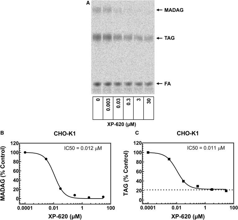
Concentration dependent inhibition of intracellular synthesis of MADAG in CHO-K1 cells by XP-620. The lipid synthesis of CHO-K1 cells was analyzed in 6-well plates when cells were at ∼90% confluence. The cells were incubated with 0.5 mM [14C]oleic acid for 1 h in the absence or presence of a series of concentrations of XP-620. The lipid products were extracted and developed in TLC plates. To calculate IC50 values, percent of MADAG or TAG formed in the presence of each concentration of XP-620 was compared with DMSO (100% control). Dose response curves were plotted and XP-620 IC50 values were calculated for MADAG or TAG using the “dose-response variable slope” model in the GraphPad Prism program. A: Phospho Image profile of intracellular synthesis of MADAG and TAG by CHO-K1 cells. B: XP-620 concentration-dependent inhibition of intracellular synthesis of MADAG in CHO-K1 cells. C: XP-620 concentration dependent inhibition of intracellular synthesis of TAG in CHO-K1 cells.

