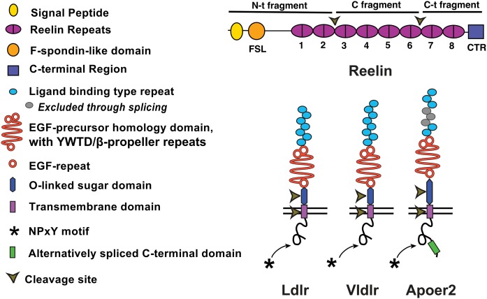
Fig. 1.
Structure of Reelin and the ApoE receptors, Ldlr, Vldlr, and Apoer2. Reelin comprises a signal peptide, an F-spondin-like domain, eight RRs that each contain a “A” and “B” parts separated by an EGF motif, and a C-terminal domain. Reelin is cleaved at two sites between RR2/RR3 and RR6/RR7. The ApoE receptors contain an extracellular ligand binding domain, EGF-precursor homology domain, an alternatively spliced OLS domain, a single transmembrane domain, and a short ICD containing one to three NPXY motifs. Apoer2 has an alternatively spliced 59 amino acid ICD.

