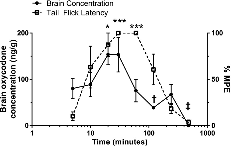
Fig. 3.
Oral oxycodone time course: antinociception and brain concentration. After a single administration of oxycodone (16 mg/kg, PO), brain oxycodone concentrations were plotted against oxycodone’s antinociceptive effect in the warm-water tail-withdrawal assay at various time points ranging from 5 to 480 minutes. All data points represent the mean ± S.E.M. from a minimum of five mice. Brain oxycodone concentrations increased during the first 20 minutes, where a plateau was observed until 30 minutes. Antinociception was slower to reach 100%MPE, which was not observed until 30 minutes and persisted until 60 minutes. Brain oxycodone concentrations at 60 minutes were much lower, measuring closer to the 5-minute time point, despite maximum antinociception. Significant observations for brain oxycodone concentrations were only noted at 120- (†P < 0.05) and 480-minute (‡P < 0.001) time points (one-way ANOVA), where concentrations were lower than all other time points. Antinociception was significantly higher at 20 (*P < 0.05), 30, and 60 minutes (***P < 0.001) compared with 5 minutes. Antinociception at 30 and 60 minutes was significantly higher compared with 240 (P < 0.001) and 480 minutes (P < 0.0001).

