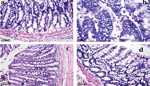
Figure 3.

Photomicrograph of Colon Tissue Section of (a): negative control rat showed normal histological structure of mucosa, (b): colon cancer bearing rat showed absence of the crypts and goblet cells of mucosa with dysplastic, hyperchromatic irregular arrangement of nuclei with hyperplasia, (c): colon cancer bearing rat treated with quercetin compound showed normal histological structure of glandular epithelium with few inflammatory cells infiltration in the lamina propria and (d): colon cancer bearing rat treated with 5-FU showed normal histological structure of mucosa.
