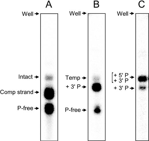
Figure 3.
Examples of gels used to monitor products from each round of primer-free genomic SELEX. (A) Isolation of primer-free DNA after restriction enzyme and alkaline treatments (Figure 1, Step 4). (B) Isolation of samples after synthesis of the 3′ primer-annealing sequence (Figure 1, Steps 6–7). Note that the amount of the templates was at least 2-fold higher than that of the primer-free DNA; the templates were intentionally reduced in radioactivity to give a lighter band on the gel. (C) Isolation of samples after ligation of the 5′ primer-annealing sequence (Figure 1, Step 9). Shown are phosphor images of 8% denaturing polyacrylamide gels. Intact, dsDNA PCR product; Comp strand, complementary strand; P-free, primer-free DNA; Temp, template DNA; + 3′P, DNA containing the 3′ primer-annealing sequence; + 5′P and + 3′P, DNA containing both 5′ and 3′ primer-annealing sequences.

