Figure 2.
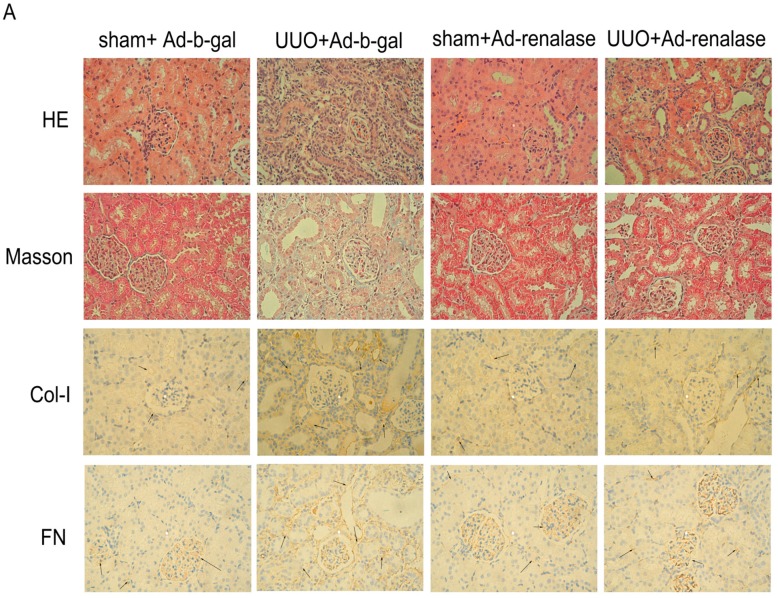
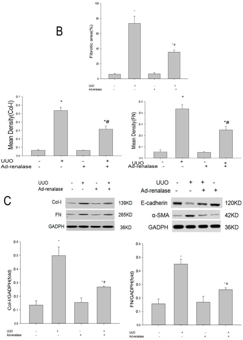
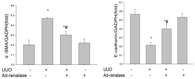
Renalase ameliorates renal interstitial fibrosis in the UUO model. (A) Kidney sections from various groups at 14 days after UUO were subjected to H&E, Masson’s trichrome staining (MTS) and immunohistochemistry. Representative micrographs showing that renalase ameliorated renal fibrotic lesions after obstructive injury. Immunohistochemistry showed that renalase reduced fibronectin and collagen I deposition in the obstructed kidney. Arrows refer to positive results. magnification: 40×; (B) Quantitative determination of renal fibrotic lesions and quantitative analysis of immunohistochemistry in various groups. Renal fibrotic lesions (defined as the percentage of the MTS-positive fibrotic area) were quantified by computer-aided morphometric analyses. That is, the fibrosis and the expression of FN and Col-I was lightened in UUO + Ad-renalase group compared to UUO + Ad-b-gal group, but was still severe compared with sham groups; (C) Western blot showed that renalase inhibited renal expression of α-SMA, fibronectin, and collagen I and increased renal expression of E-cadherin in the ligated kidney at 14 days after UUO. Results are presented as percentages of control values and are the means ± SD of five animals per group. * p < 0.05, compared with the sham + Ad-b-gal group; # p < 0.05, compared with the UUO + Ad-renalase group.


