Figure 3.
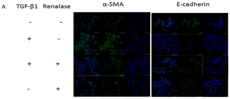
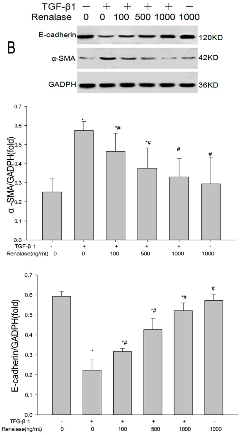
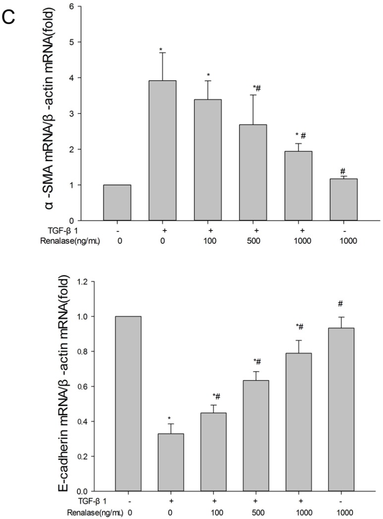
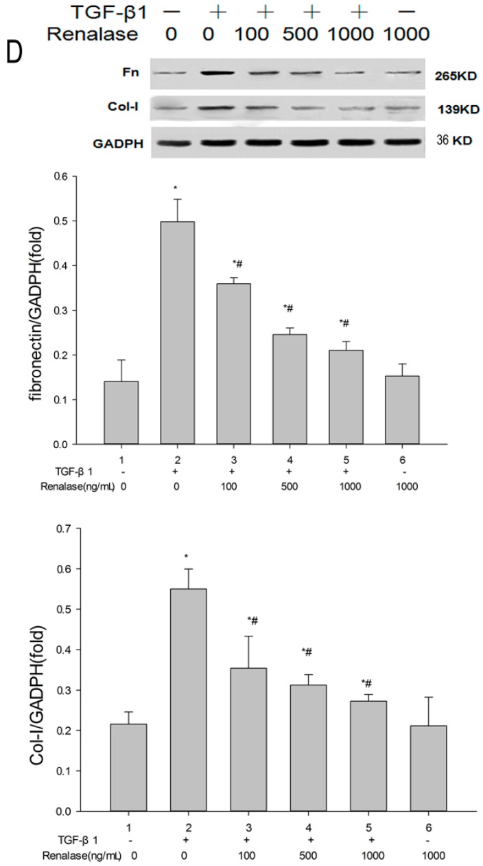
Renalase blocks transforming growth factor-β1 (TGF-β1)-mediated epithelial to mesenchymal transition (EMT) in vitro. Human proximal tubular epithelial cells (HK-2) were treated with 2 ng/mL TGF-β1 in the presence or absence of various concentrations of renalase as indicated for 48 h. (A) Immunofluorescence showed that renalase (1000 ng/mL) abolished TGF-β1-induced α-smooth muscle actin (α-SMA) assembly and preserved E-cadherin integrity; magnification: 120×; (B,D) Western blotting demonstrated that renalase (100, 500 and 1000 ng/mL) reversed the increased expression of α-SMA, collagen-I, and fibronectin, and decreased expression of E-cadherin in a dose-dependent manner; (C) reverse transcription (RT)-PCR revealed that renalase (100, 500 and 1000 ng/mL) reversed the increased Messenger RibonucleicAcid (mRNA) expression of α-SMA and decreased mRNA expression of E-cadherin in a dose-dependent manner. Results are presented as percentages of control values after normalization to GADPH and are the means ± SD of three independent experiments. * p < 0.05, compared with control groups; # p < 0.05, compared with TGF-β1-stimulated groups; n = 3 per group.


