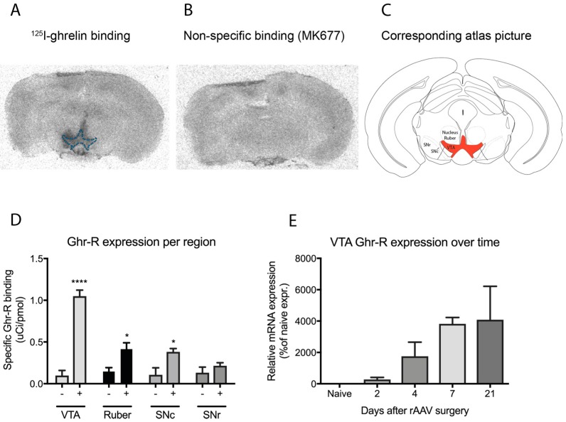
Figure 1.
(A) Representative image of a brain section containing the ventral tegmental area (VTA; outlined in blue) from a ghrelin receptor (Ghr-R)VTA mouse after autoradiography, showing specific binding of 125I-ghrelin to the Ghr-R; (B) Representative image of a brain section containing the VTA from a Ghr-RVTA mouse after autoradiography, showing binding of non-labeled MK677, a Ghr-R agonist, to visualize non-specific binding of 125I-ghrelin; (C) The corresponding atlas picture with the VTA outlined in red, adapted from Paxinos & Keith 2001, figure 60 [36]; (D) Ghr-R expression of Ghr-RVTA mice (+) and Ghr-R knockout (KO) control mice (−) in different regions of the midbrain, namely the VTA, nucleus ruber, substantia nigra pars compacta (SNc) and substantia nigra pars reticulata (SNr), n = 7; (E) Ghr-R expression in the VTA of Ghr-RVTA mice at different time points after rAAV-mediated re-expression of the receptor in the VTA of Ghr-R KO mice, n = 2. * p ≤ 0.05, **** p ≤ 0.0001. All data are expressed as mean ± SEM.

