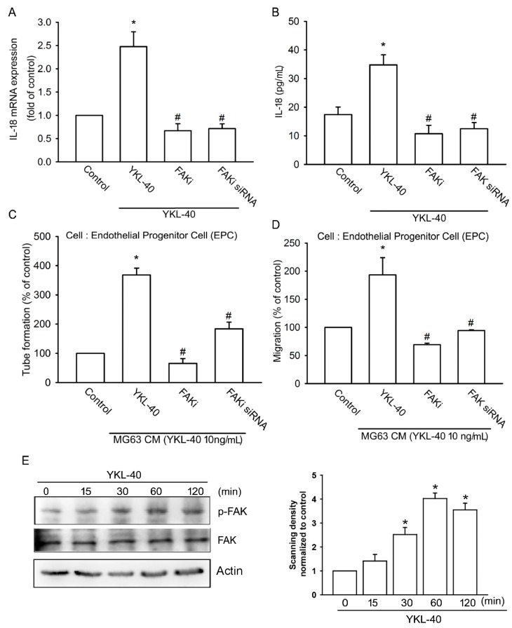
Figure 3.
The focal adhesion kinase (FAK) signaling pathway regulates YKL-40-induced increases in IL-18 expression. (A,B) MG-63 cells were pretreated with a FAK inhibitor (10 μM) or transfected with FAK siRNA for 24 h, then stimulated with YKL-40 for 24 h. IL-18 expression was examined using qPCR and ELISA assays (n = 4 per group). (C,D) CM was collected and applied to EPCs. Capillary-like structure formation and cell migration of EPCs was examined by tube formation and Transwell assay (n = 5 per group). (E) MG-63 cells were treated with YKL-40 for indicated time intervals, and FAK phosphorylation was examined by Western blotting. FAK phosphorylation in each independent experiment was quantified by densitometry in right panel (n = 3 per group). Results are expressed as the mean ± S.E. * p < 0.05 compared with control. # p < 0.05 compared with the YKL-40-treated group.

