Figure 3.
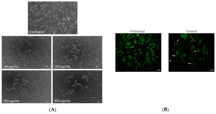
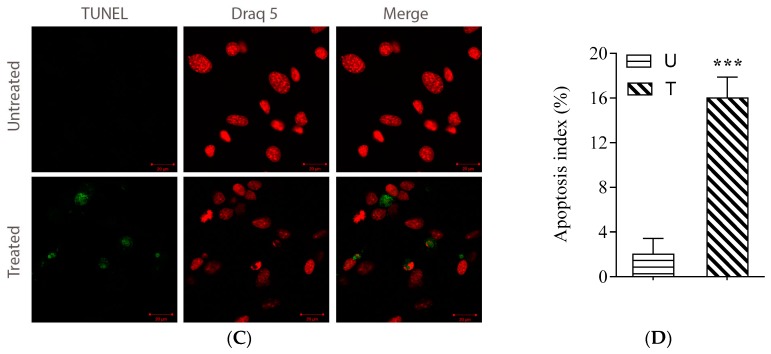
Induction of apoptosis by AEE extract. Contrast-phase microscopy on B16-F10 murine melanoma cells was untreated and treated with AEE, in coverslides, for 24 h (A). Fluorescence microscopy by dual staining AO/EB (acridine orange/ ethidium bromide) of B16-F10 cells. Untreated cells and cells with intact membrane appeared colored green, being permeable for EB. B16-F10 cells treated AEE (250 μg/mL) labeled with both dyes simultaneously, apoptotic cells appeared orange (indicated with white arrows) (B). TUNEL assay for apoptosis detection. B16-F10 cells were treated with AEE (250 μg/mL) for 24 h and analyzed for apoptosis using an in situ cell death kit ApopTag® Red in situ Apoptosis Detection kit (Chemicon, Millipore, Billerica, MA, USA). TUNEL-positive cells are shown as green fluorescence; normal nuclei are stained red with Draq5 (C). Quantification of TUNEL assay. Data are presented as percentage of TUNEL-positive cells per 1000 cells; U-untreated cells; T-AEE treated cells (D).


