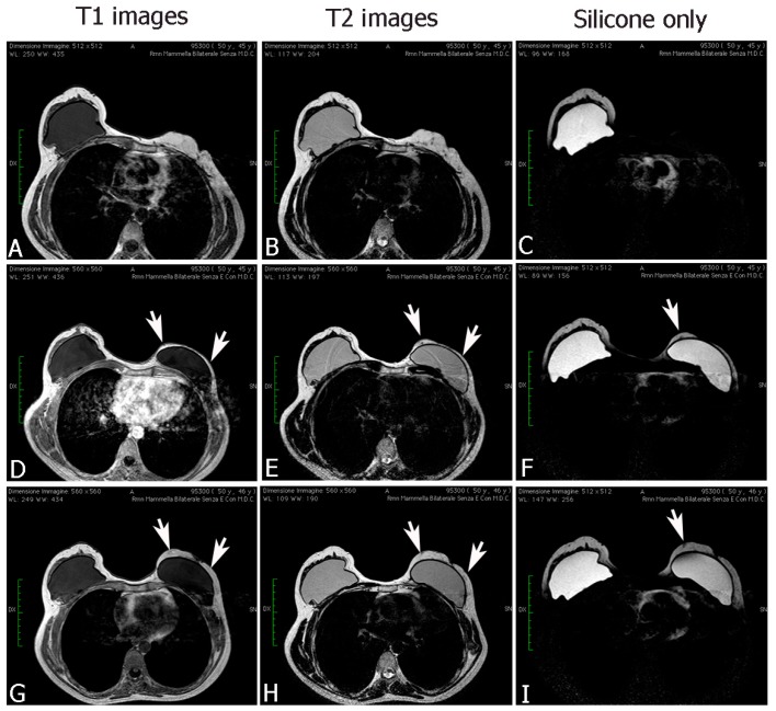
Figure 7.
MRI figures, after fat transplantation to the breast, showed changes in volume, in thickness, and in fat distribution in the spaces (G–I) with respect to a previous treatment (D–F), and with respect to the pre-operative treatment (A–C). The fat grafting site was visible on MRI scans, with an identical signal of adjacent fat. The injected fat appearance is that of normal breast fat: hyperintense on T1- and T2-weighted sequences. On T1-weighted fat suppression images, the injected fat showed a hypointense normal fat signal. No signs of necrosis were presented on MRI.

