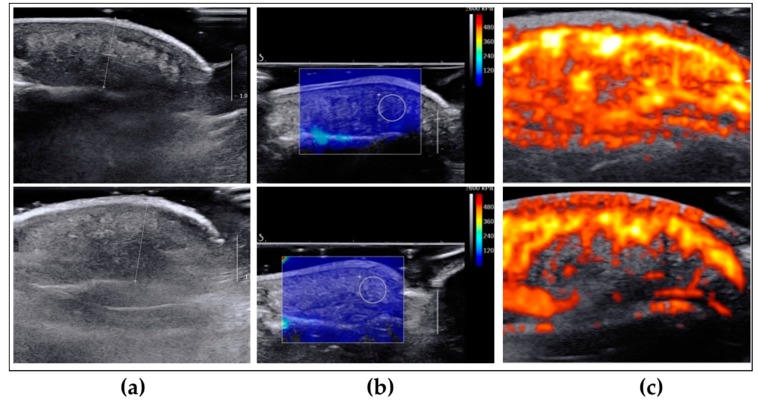
Figure 2.
Representative echo-doppler images of regenerated (upper row) and un-injured collateral (lower row) fingertips. (a) Ultrasound b-mode imaging for the measurement of the pulp thickness; (b) b-mode imaging with superimposed share wave mapping for the measurement of the pulp elasticity. The white circle has a diameter of 5 mm; (c) b-mode imaging with superimposed power Doppler for the measurement of vascularity.

