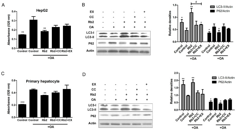
Figure 8.
Inhibition of sirt1 and AMPK blocked Rb2-induced hepatic autophagy. HepG2 cells (A,B) and primary mouse hepatocytes (C,D) were pretreated with 50 µmol/L Rb2 for 4 h in the presence or absence of the sirt1 inhibitor EX-528 (EX) and the specific AMPK inhibitor Compound C (CC), and then subjected to OA (1 mmol/L for HepG2 and 2 mmol/L for primary mouse hepatocytes) exposure for 12 h. For lipid content determination, intracellular TG were stained by Oil red O (ORO). ORO was then eluted with isopropanol and the optical absorbance of the eluate was measured at 520 nm (n = 3). Data are expressed as mean ± SE from three independent experiments. * p < 0.05, ** p < 0.01 and *** p < 0.001 compared with the group that was pretreated with vehicle (DMSO) prior to OA exposure. # p < 0.05 compared between the two indicated groups. For autophagic activity evaluation, levels of LC3-II and P62 were detected and quantified by Western blotting analysis.

