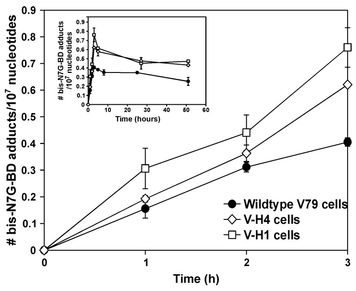
Figure 4.
Time course for the formation (main panel) and repair (insert) of bis-N7G-BD cross-links in V79 (circles), V-H1 (squares), and V-H4 (diamonds) cells. Cells were exposed to 15 µM DEB for 1, 2, or 3 h. At the indicated times, chromosomal DNA was isolated, and bis-N7G-BD adduct levels were quantified by HPLC-ESI+-MS/MS as described in the Methods section. Results represent average ± the standard error of the mean, n = 2. Insert: ICL dynamics following DEB removal. Following treatment, DEB was removed and replaced with fresh growth media. DNA was isolated, and bis-N7G-BD adduct levels were determined as described above.

