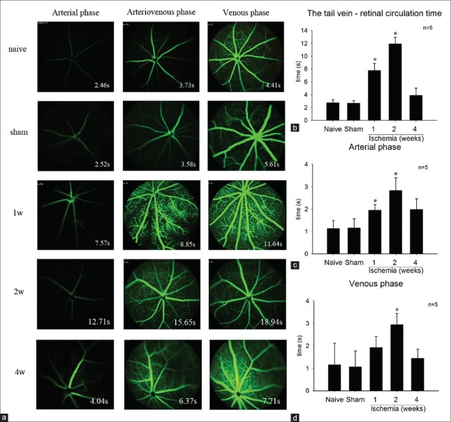
Figure 3.
(a) The fundus fluorescein angiography of each group at different phase. (b) The tail vein-retinal circulation time was significant prolonged in 1- and 2-week group. However, it was no significant difference between the 4-week group and the sham or naive group. (c) The arterial phase was significant prolonged in the 1- and 2-week group compared with that of the sham or naive group. And, there was no significant difference between the 4-week group and the control group. (d) The venous phase significant prolonged in the 2-week group. *P < 0. 01 compared with naive group.

