Figure 1.
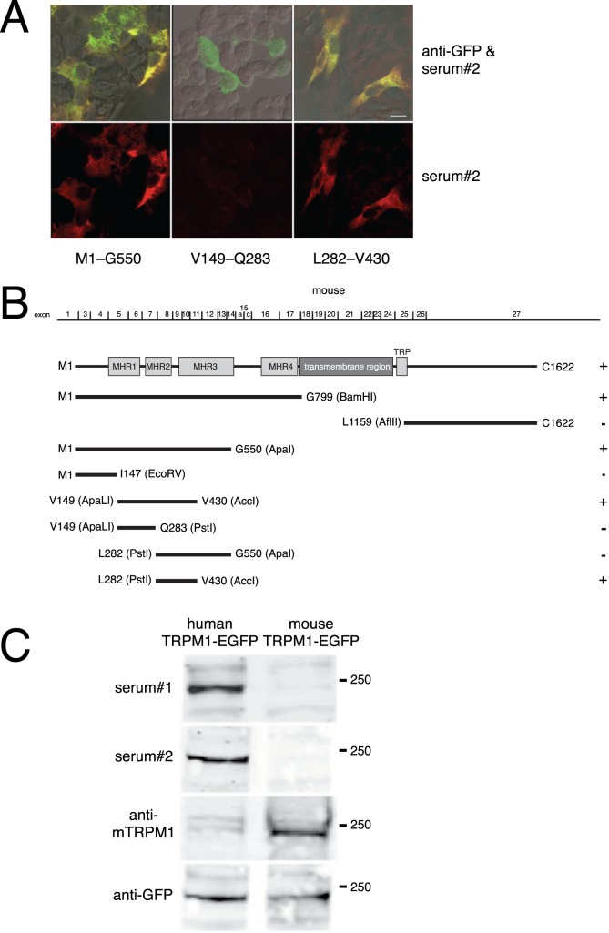
The MAR epitope is encoded by mouse TRPM1 exons 8 through 11. To map the MAR epitope, HEK cells were transfected with a series of EGFP–mouse TRPM1 deletion constructs and tested for immunofluorescence with MAR serum. (A) Top row: superimposition of GFP (green) and MAR serum 2 (red) immunofluorescence, with colocalization appearing yellow. Bottom row: MAR serum immunofluorescence alone. Scale bar: 10 μm. (B) Diagram of the mouse TRPM1 cDNA deletion constructs. Exon 2, which is alternatively spliced and encodes an alternative N-terminus, is not present in the plasmid constructs used. The first and last amino acids encoded by each construct are indicated. Positive immunofluorescence with MAR serum was graded as positive (+), or negative (−). MHR: TRPM homology regions.26 (C) HEK293 cells were transfected with plasmids encoding either mouse TRPM1-EGFP or human TRPM1-EGFP and then Western blotted with either MAR serum 1, MAR serum 2, an antibody to mouse TRPM1, or an antibody to GFP.

