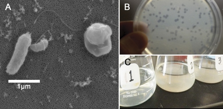
Figure 1. Halobacteriovorax predation of Vibrio spp.
(A) Micrograph of the pathogen, Vibrio coralliilyticus BAA450 being attacked by Halobacteriovorax and rounded V. coralliilyticus bdelloplast (right) with Halobacteriovorax inside (B) double layer plate showing freshly lysed plaques on a lawn of V. coralliilyticus cells (C) Overnight liquid cultures of (1) a co-culture of Halobacteriovorax and V. fortis, (2) V. fortis and 0.2 µm filtrate from Halobacteriovorax culture, and (3) V. fortis alone.

