Figure 5.
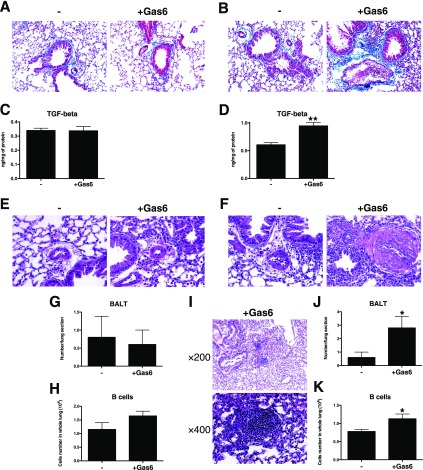
Exogenous Gas6 uniformly induced fibrosis, vascular smooth muscle hypertrophy, and B cell accumulation. Representative Masson trichrome–stained lung tissue sections from the low-dose (2 μg) Gas6 (A) and high-dose (7 μg) Gas6 groups (B) at Day 28 after conidia challenge. TGF-β transcript and protein levels in the low-dose (C) and high-dose (D) Gas6-treated groups at Day 28 after conidia challenge. Vascular wall hypertrophy was detected in the low-dose Gas6 (E) and high-dose Gas6 (F) groups at Day 28 after conidia challenge. The number of bronchus-associated lymphoid tissue (BALT) follicles was not changed in the low-dose Gas6-treated lung (G), but the number of B cells in the BALT from this group was increased (H). Expanded BALT was observed in histological sections from the high-dose Gas6-treated group (I), and the number of follicles (J) and the number of B cells in these follicles (K) were significantly increased compared with the control group. Results are expressed as the mean ± SEM for n = 5 per group. *P < 0.05 and **P < 0.01 versus control (indicated as -).

