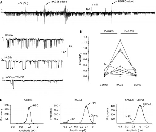
Figure 4.
hAGEs regulate ENaC activity via oxidant signaling in rat primary alveolar type 2 cells. (A) Continuous cell-attached single-channel recording of a primary alveolar type 2 cell accessed from a lung slice preparation. Arrow represents the closed (c) state, with downward deflections from the arrow representing inward Na+ channel openings (−40 mV [−Vp] holding potential). Enlarged portions of the representative recording represent control, hAGE treatment, and TEMPO, a SOD mimetic, conditions. (B) Results from eight independent observations shown on dot plot, with ENaC activity on the y axis. Challenge with hAGEs increased ENaC NPo from 0.12 ± 0.05 to 0.53 ± 0.16 (P = 0.025), and the addition of TEMPO decreased ENaC NPo to 0.10 ± 0.03 (P = 0.013). (C) Point amplitude histograms demonstrate that a challenge with hAGEs increase HSC channel and NSC channel activity in isolated alveolar type 2 cells.

