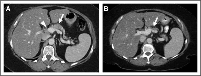
Fig 2.
(A and B) Axial computed tomography (CT) images of the abdomen (A) before and (B) after neoadjuvant chemotherapy. (A) An initial diagnostic abdominopelvic CT scan demonstrated a hypodense mass in the proximal pancreatic body (arrow), with abutment of the portal vein–superior mesenteric vein confluence (arrowhead). (B) A follow-up abdominopelvic CT scan after 12 cycles of modified FOLFIRINOX (fluorouracil, folinic acid, irinotecan, and oxaliplatin) demonstrated a decrease in the size of the pancreatic body mass (arrow).

