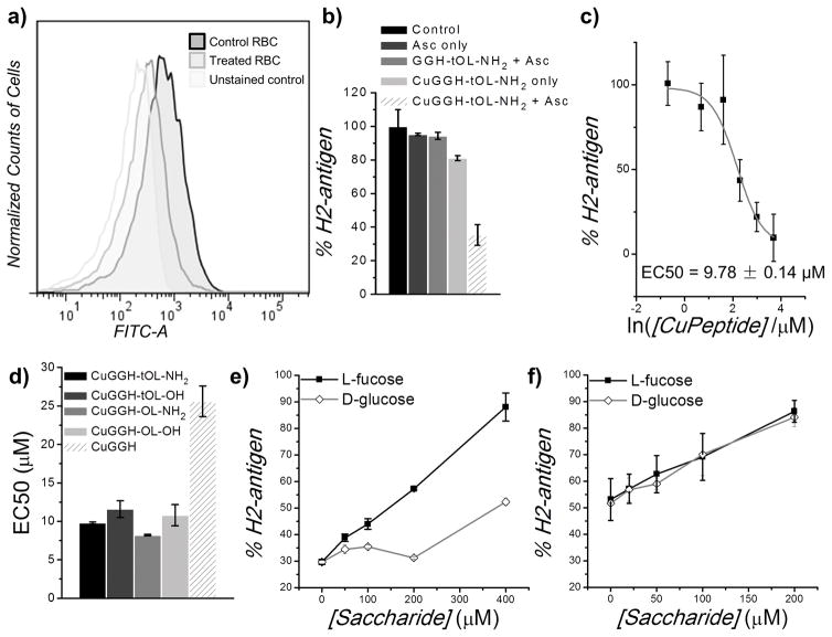
Figure 2.
Removal of H2-antigen from erythrocytes. a) Flow cytometric analysis of H2 antigen on the surface of human red blood cells (RBC). After erythrocytes were incubated with 20 μM CuGGH-tOL-NH2 and 200 μM ascorbate for 4 h, immunofluorescent staining was performed with a FITC-labelled anti-H2 antibody. b) Relative amount of H2 antigen on erythrocytes, after incubation with or without peptide or ascorbate. Solutions containing 200 μM ascorbate and 20 μM CuGGH-tOL-NH2 were used. c) Relative amount of H2 antigen on erythrocytes after incubation with various amount of CuGGH-tOL-NH2. d) The half-maximal effective concentration (EC50) for H2-antigen removal by artificial fucosidases in the presence of 200 μM ascorbate. e) and f) Inhibitory effect of saccharides. Human erythrocytes were incubated with 20 μM CuGGH-tOL-NH2 (e), or CuGGH (f), with the indicated concentration of saccharides, and 200 μM ascorbate.

