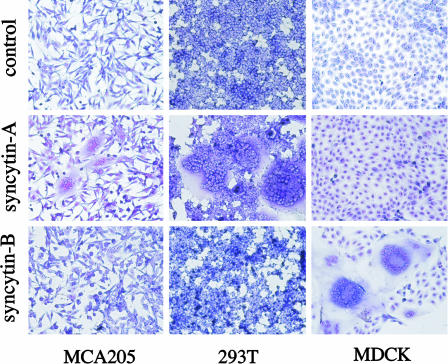
Fig. 6.
Syncytin-mediated cell–cell fusion. Shown is syncytia formation by the syncytin-A and -B envelope genes. The indicated cells were transfected with expression vectors for syncytin-A or -B, or a negative control (syncytin-A in antisense orientation) and were stained with May–Grünwald and Giemsa 24–48 h after transfection.

