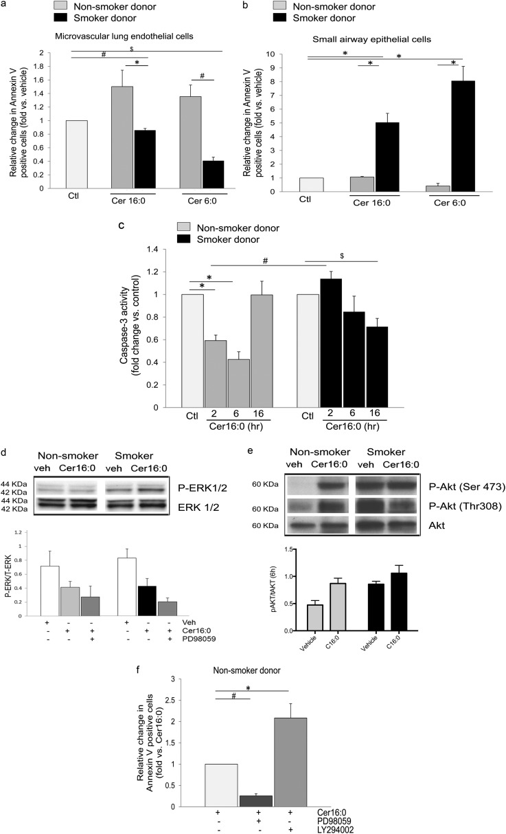
Figure 2.
Apoptosis and survival responses of human lung microvascular endothelial cells to Cer16. (a and b) Apoptosis measured by annexin V/propidium iodide staining and expressed as fold increase in positive cells versus control vehicle in human lung microvascular endothelial cells (a) compared with human small airway epithelial cells (b), both isolated from either nonsmokers (gray bars) or smokers (black bars). Cells were treated with Cer16 or Cer6 (10 μM; 6 h), or with vehicle (PEG 2,000); mean + SEM (n = 4; *P < 0.05, #P = 0.006, and $P < 0.001). (c) Caspase-3 activity of human lung microvascular endothelial cells treated Cer16 (10 μM) or vehicle (control [Ctl]) for the indicated time. Mean + SEM. (n = 9; *P < 0.05, #P = 0.006, and $P = 0.05. (d) Activated and total extracellular signal–regulated kinase (ERK) 1/2 measured by Western blot in lung microvascular endothelial cells (LMVECs) lysates from nonsmoker and smoker donors after treatment with Cer16 (10 μM) or vehicle (veh) for 6 hours (upper panel, Western blot) and 16 hours (lower panel, densitometry). Mean + SEM; n = 4. (e) Activated and total Akt in LMVEC lysates treated with either Cer16 (10 μM) or vehicle for 6 hours (upper panel showing Western blot and lower panel showing densitometry). Mean + SEM (n = 4; two-way ANOVA showing P < 0.05 for both Cer treatment and smoking status). (f) Effect of ERK1/2 or Akt inhibition on LMVEC apoptosis measured by annexin V/PI staining in cells from nonsmoker donors. Cells were pretreated (1 h) with either PD98059 (10 μM) or LY294002 (30 μM) followed by Cer16 (10 μM, 2 h). Results are expressed as fold change versus Cer16. Mean + SEM (n = 3; *P = 0.01, #P < 0.0001). P-Akt, phosphorilated serine/threonine-specific protein kinase; P-ERK, phosphorilated extracellular signal–regulated kinase; Ser, serine; Thr, threonine.

