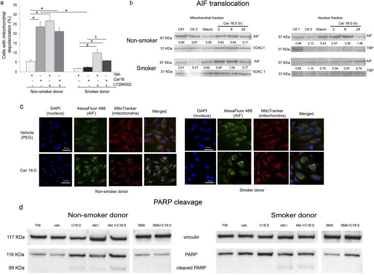
Figure 3.
Mitochondria changes in human lung microvascular endothelial cells treated with Cer16. (a) Mitochondria depolarization in cells treated with Cer16 (10 μM, 6 h) or vehicle (Veh) and effect of Akt inhibitor LY294002 (30 μM, 1 h pretreatment). Mean + SEM (n = 3; *P < 0.05, #P < 0.001, $P = 0.05). (b and c) Apoptosis-inducing factor (AIF) translocation from mitochondria to nucleus detected by Western blot (b) in respective cellular subfractions of cells treated with Cer16 (10 μM; for the indicated time in hours) or its Veh (PEG 2000; Ctl1) compared with staurosporine (Stauro; 0.2 μM, 2 h) or its Veh (Ctl2). Loading controls for each subcellular fraction, voltage-dependent anion channel (VDAC1) and TATA-binding protein (TBP), were detected in the lower lanes. Densitometry of AIF expression normalized by loading control is indicated numerically in between lanes. (c) Representative fluorescence micrographs of cells immunostained for AIF (FITC-labeled). Mitochondria are stained in red (MitoTracker Red) and nuclei in blue with 4′,6-diamidino-2-phenylindole (DAPI) after treatment with Cer16 (10 μM) or Veh; scale bar, 50 μm. (d) Detection of cleaved poly(ADP-ribose) polymerase (PARP) in total protein lysates of cells: untreated, grown in regular full serum–containing media (FM); or treated with Veh or Cer16 (10 μM, 6 h), and either Akt inhibitor (30 μM LY294002; 1 h pretreatment) or autophagy inhibitor, 3-methyladenine (3-MA; 5 mM, 1 h pretreatment). Vinculin expression was used as loading control.

