Figure 4.
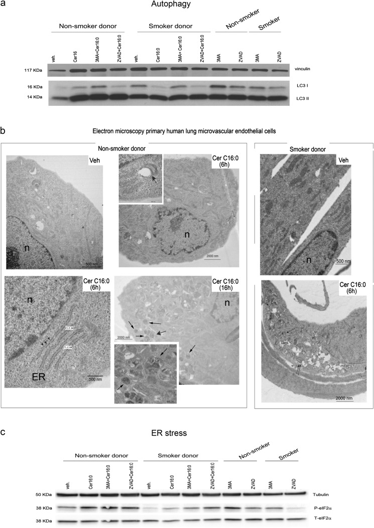
Autophagy and endoplasmic reticulum (ER) stress in human lung microvascular endothelial cells treated with Cer16. (a) Western blot of microtubule-associated protein-1 light-chain 3 (LC3) -I and LC3-II and vinculin (as loading control) in cells treated with Cer16 (10 μM, 16 h) or Veh, and effect of general caspase inhibitor, known commercially as ZVAD-fmk (0.1 mM, 1 h pretreatment) or autophagy inhibitor 3-MA (5 mM, 1 h pretreatment). (b) Representative electron microscopy images of cells after treatment with Cer16 or Veh for 6 or 16 hours. Noted are: nuclei (n), ER swelling (arrowheads, lower panel), autophagosomes (magnified in lower panel inset, arrows), and autophagosome–lysosome fusion (magnified in upper panel inset, arrow). (c) Western blot of phospho- and total eukaryotic translation initiation factor 2a (eIF2α) in cells similarly treated as in (a). Cells were treated with Cer16 or Veh control (PEG 2,000; 10 μM, 16 h) and pretreated with ZVAD-fmk (0.1 mM, 1 h) or 3-MA (5 mM, 1 h). β-tubulin was used as loading control.

