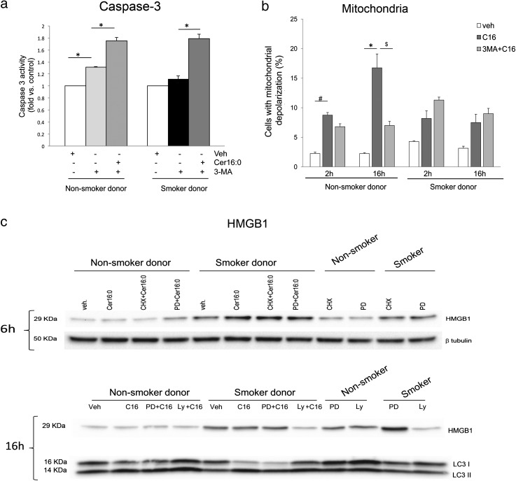
Figure 5.
Autophagy and apoptosis interactions in human lung microvascular endothelial cells treated with Cer16. (a and b) Apoptosis measured by caspase-3 activity (a) or mitochondrial depolarization (b) in cells treated with Cer16 (10 μM; 2 hours in [a] and indicated time in [b]) or Veh, and effect of autophagy inhibitor, 3-MA (5 mM, 1 h pretreatment). Mean + SEM (n = 4, *P < 0.05). (c). Levels of high-mobility group box 1 (HMGB1) or LC3-I and LC3-II measured by Western blot in cells treated with Cer16 (10 μM, for the indicated time) or Veh and either protein synthesis inhibitor, cycloheximide (CHX; 1 μg/ml, 1 h pretreatment), ERK1/2 inhibitor, PD98059 (PD; 50 μM, 1 h pretreatment), or Akt inhibitor LY294002 (LY; 30 μM, 1 h pretreatment).

