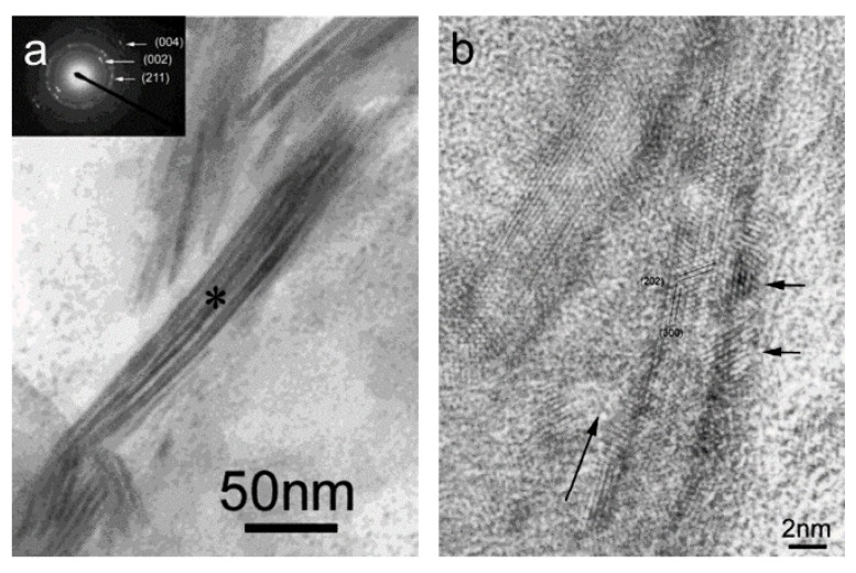
Figure 2.
(a) Higher magnification of the mineralized collagen fibrils. Insert is the selected area electron diffraction pattern of the mineralized collagen fibrils. The asterisk is the center of the area and the diameter of the area is about 200 nm. (b) High-resolution transmission electron microscopy (HR-TEM) image of mineralized collagen fibrils. Long arrow indicates the longitude direction of the collagen fibril. Two short arrows indicate two hydroxyapatite (HA) nanocrystals. Reprinted with permission from reference [44]. Copyright 2003 American Chemical Society.

