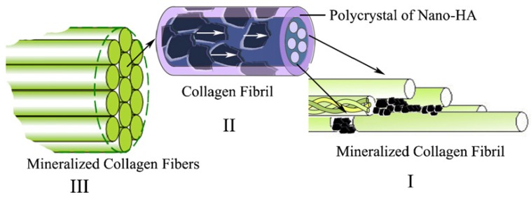
Figure 3.
The hierarchical structure of the self-assembled mineralized collagen. (In I, the first level, the organization of the collagen molecules with the nano-sized HA crystals formed initially in the gap zones between the collagen fibrils; in II, the second level, showing the organization of collagen fibrils with respect to HA crystals, the HA crystals are platelet-like and grow on the surface of these fibrils in such a way that their c-axes are oriented along the longitudinal axes of the fibrils, as indicated by the white arrows in the figure; in III, the third level, showing the organization of the mineralized collagen fibrils, a number of mineralized collagen fibrils align parallel to each other to form mineralized collagen fibers. Reprinted with permission from reference [1]. Copyright 2007 Elsevier.)

