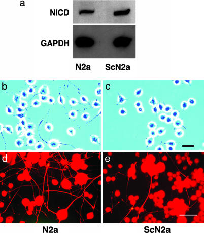
Fig. 3.
ScN2a cells have a higher concentration of NICD and fewer cells with neurites compared with N2a cells. (a) Western blots show ≈2× as much NICD in ScN2a cells (cleaved Notch-1 Val-1744 antibody). GADPH was used to normalize the data. (b and c) Phase-contrast microscopy shows that many N2a cells grew neurites that are more than two cell diameters in length (b); in contrast, most processes of ScN2a cells are less than two cell diameters in length (c). (d and e) Fluorescence immunohistochemistry shows that the cell bodies and long neurites of both N2a (d) and ScN2a cells (e) are immunopositive for the high molecular-weight neurofilament protein, NF200. N2a cells grew far more NF200-immunopositive neurites than ScN2a cells. (Bars = 90 μm; bar in c applies to b; bar in e applies to d.)

