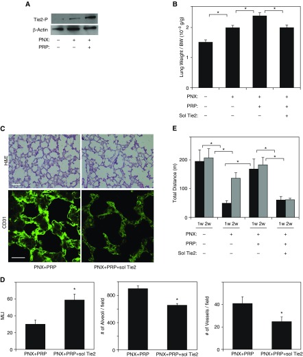
Figure 4.
PRP extract induces angiogenesis and compensatory lung growth in mouse lung after PNX through Ang1–Tie2 signaling. (A) Immunoblots showing tyrosine-phosphorylated Tie2 and β-actin protein levels in the right lung cardiac lobe of control mouse, mouse 7 days after PNX, or in combination with treatment with PRP extract for 7 days after PNX. (B) Graph showing the ratio of weight of the right lung cardiac lobe to mouse body weight after PNX treated with PRP extract or in combination with soluble (sol) Tie2 receptor for 7 days (n = 7; mean ± SEM). *P < 0.05. Protein concentration–matched mouse serum and control vehicle are used as a control. (C) H&E–stained mouse right lung cardiac lobe treated with PRP extract or in combination with sol Tie2 for 7 days after PNX (top; scale bar, 50 μm). Immunofluorescence micrographs showing CD31-positive blood vessels in right lung cardiac lobe of mouse treated with PRP extract or in combination with sol Tie2 for 7 days after PNX (bottom; scale bar, 20 μm). (D) Graphs showing the quantification of alveolar size (MLI, left), the alveolar number (middle), and the vessel number of CD31-positive endothelial cells (right) in the right lung cardiac lobe after each treatment (n = 7; mean ± SEM). *P < 0.05. (E) Exercise capacity of mice 7 and 14 days after PNX or in combination with treatment with PRP extract and/or sol Tie2 receptor after PNX for 7 and 14 days assessed by a total running distance using a rodent treadmill exercise protocol (n = 7; mean ± SEM). *P < 0.05.

