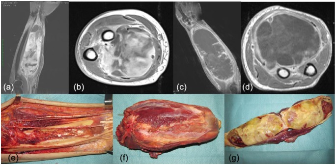
Figure 2.
A 58-year-old man was diagnosed with a 6 × 20 cm mass in his left volar forearm. Percutaneous core needle biopsy revealed a high-grade myxofibrosarcoma (a, b) [contrast-enhanced magnetic resonance imaging (MRI), TW1 weighted sequences]. This patient was treated with three cycles of epirubicin (120 mg/m2) and ifosfamide (9000 mg/m2) and concomitant radiotherapy (50 Gy in 25 fractions). MRI showed an increase in tumour dimension and a strong reduction of tissue contrast enhancement, suggesting a tissue response (c, d). Surgery involved a wide excision of the posterior forearm (e). The tumour was resected together with the median nerve which was completely surrounded by the tumour (f, g). The pathology report showed significant presence of necrosis (70% of the tumour mass) and limited residual tumour (30%).

