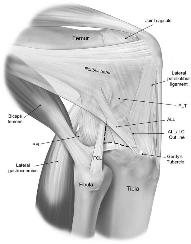
Figure 2.

Lateral view of a right knee illustrating the anterolateral corner structures. The dashed line shows where the anterolateral ligament and lateral capsule (ALL/LC) were sectioned deep to the superficial layer of the iliotibial band (ghosted). The ALL/LC was released from the anterior margin of the fibular collateral ligament (FCL) extending 7 cm anteromedial to the center of ALL tibial attachment. The ALL/LC was released as one unit. PFL, popliteofibular ligament; PLT, popliteus tendon.
