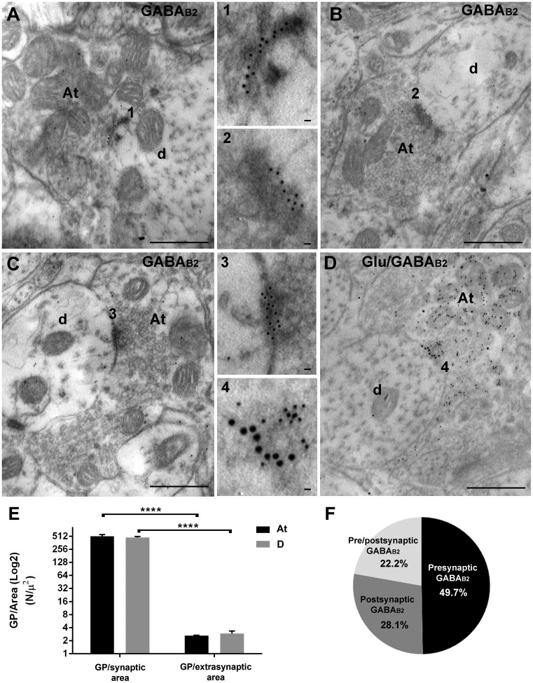
Figure 2.
Localization of GABAB2 at axo-dendritic synapses in laminae III-IV. (a) A GABAB2-immunoreactive At makes a synapse with an unlabeled dendrite (d). GPs are specifically distributed along the presynaptic membrane (insert number 1). (b) A unlabeled At makes a synapse with a GABAB2-immunoreactive dendrite (d). GPs are distributed along the postsynaptic density (insert number 2). (c): A GABAB2-immunoreactive At makes a synapse with a GABAB2-immunoreactive dendrite (d). GPs are scattered over both the pre- and postsynaptic densities (insert number 3). (d) A glutamate + GABAB2-immunoreactive At makes a synapse with an unlabeled dendrite (d). Glutamate-IR is characterized by 10 nm GPs, scattered all over the At, while GABAB2 is evidenced by 20 nm GPs specifically distributed along the presynaptic membrane (insert number 4). (e) Bar chart showing GPs densities (GP/Area, N/µ2) at synaptic and extrasynaptic sites of (At, black bars) and dendrites (D; gray bars).No statistical difference was shown between At and D for both GP/synaptic area (two-way ANOVA, not repeated measures, p = 0.5725) and GP/extrasynaptic area (two-way ANOVA, not repeated measures, p = 0.5725). GPs at the synapse were significantly different from GPs at extrasynaptic sites in both At and D (two-way ANOVA, not repeated measures followed by Bonferroni multiple comparisons test, ****p < 0.0001). (f) pie chart showing the results of quantitative analysis on the percentage of expression of GABAB2 at presynaptic sites (black; 49.7%), postsynaptic sites (dark gray; 28.1%), or both (light gray; 22.2%). Scale bar: 500 nm; inserts: 20 nm. GABA: γ-aminobutyric acid; At: axon terminal; D: dendrite.

