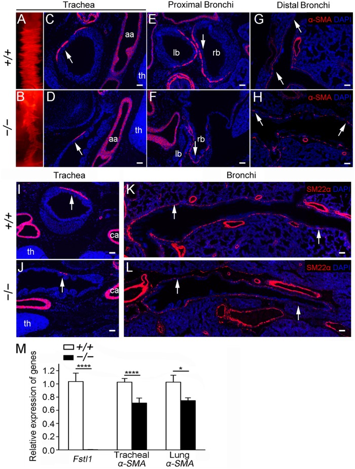
Fig 3. Loss of Fstl1 led to abnormal tracheal and bronchial SM formation in E18.5 embryos.
(A, B) α-SMA whole-mount staining revealed an extremely attenuated α-SMA signal in Fstl1−/− trachea. Trachea (C, D), proximal bronchi (E, F, the sections at the points where the tracheas split into the left and right main bronchi) and distal bronchi (G, H) sections stained for α-SMA confirmed reduced α-SMA expression (arrows). (I, J) Trachea sections of similar planes, as indicated by the common carotid artery and thymus, stained for SM22α revealed reduced SM cells in Fstl1−/− trachea. (K, L) Stitched images showed airway SM defects from proximal bronchi to distal bronchi in Fstl1−/− lung. (M) qRT-PCR analysis of the expression of Fstl1 and α-SMA in E18.5 tracheas and lungs (n = 5 per group). aa, arch of the aorta, th, thymus, ca, common carotid artery, lb, left main bronchus, rb, right main bronchus. *, P < 0.05; ****, P < 0.0001. Error bars indicate mean ± SEM. Scale bars, A, B, 200 μm; C-L, 50 μm.

