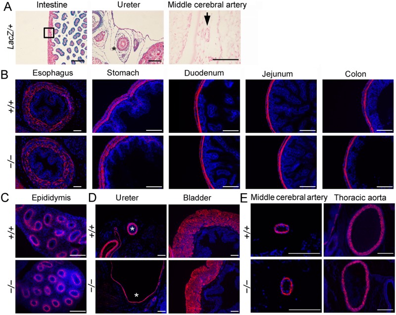
Fig 5. LacZ staining and immunostaining for α-SMA on transverse sections of other organs.
(A) LacZ staining of intestine, ureter and middle cerebral artery sections. (B) Immunostaining for α-SMA on transverse sections of E18.5 esophagus, stomach, duodenum, jejunum and colon. (C) α-SMA staining of epididymis showed normal SM differentiation in ductus epididymis. (D) α-SMA immunostaining to examinate the formation of SM of ureter and bladder. Asterisks indicate the SM layer of ureter. (E) Immunostaining for α-SMA on middle cerebral artery and thoracic aorta sections. Scale bars, 100 μm.

