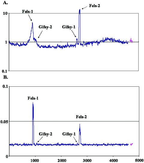
FIG. 2.
Fels phage particles released after mitomycin C treatment. LT2 cells growing in Luria-Bertani medium were treated with mitomycin C for 5 h. Subsequently, cells were separated from the supernatant, and phage were harvested from the supernatant fraction by PEG precipitation. DNA was isolated from both the cells and the supernatants. (A) LT2 cells 5 h after treatment with mitomycin C. The y axis indicates the ratio of the DNA content of treated cells to the DNA content of cells before treatment on a log10 scale. (B) Phage DNA harvested from LT2 supernatants 5 h after treatment with mitomycin C. The y axis indicates the contribution of every gene to the total signal of all genes represented on the microarray (expressed as percentages).

