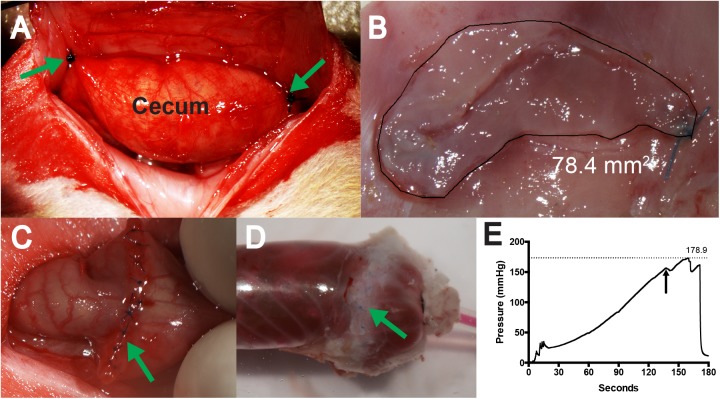
Fig 1. Experimental methods.
A. To create the cecal hinge, the abraded cecum was stitched in two places (arrows) to appose a 1 cm X 2 cm area where the peritoneum had been removed. B. Quantification of the primary adhesion. The free cecum was cut from the adhesion area, the contents removed, and the edges trimmed. The adhesion was outlined and the area recorded. C. Completed strictureplasty surgery. D. The intestinal segment 7 days following strictureplasty was instrumented and inflated until suture failure. The sutures can be seen beneath the fatpad adhesion that encased the suture line. E. Sample trace of intraluminal pressure. The first dip (arrow) was associated with mesothelial splitting at a different site from the suture line. This sample withstood a pressure of 179 mmHg.

