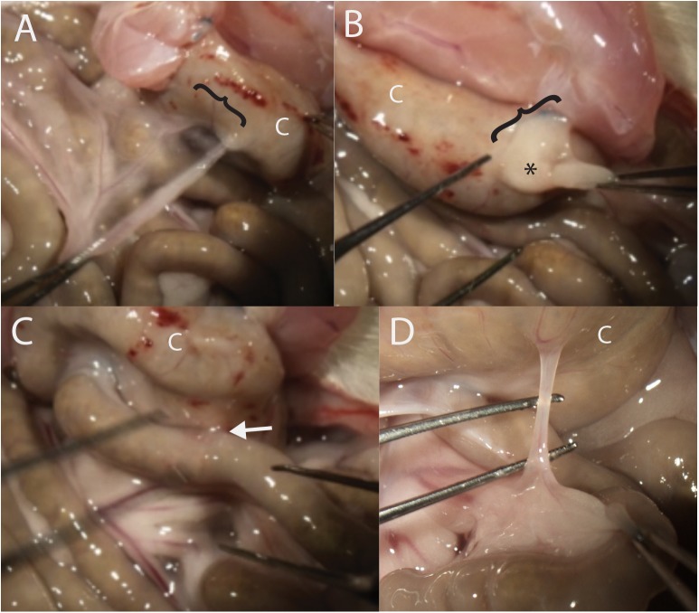
Fig 2. Representative postoperative adhesions seen in the cecal hinge model.
Images A-C were taken from the same rat in the 4-day treatment group. A. Greater omentum to cecum (C, in all panels), the extent indicated by a bracket. B. Left adnexal fatpad (*) is adherent to the cecum. In this rat there was no primary adhesion, which can be appreciated by the fold in the tissue between the abdominal wall and the cecum. C. Cohesive adhesion between the small intestine and the cecum (arrow). D. Band-like adhesion between the mesentery of the small intestine and the cecum.

