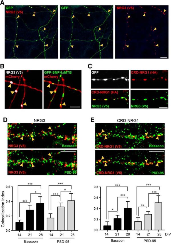
Figure 8.

NRG3 accumulates on axonal varicosities, some of which are bona fide glutamatergic terminals. A, Hippocampal neurons were cotransfected with NRG3/V5 in pDESTDV3 and GFP. The representative low-magnification image shows GFP-positive processes with V5 signal accumulating at varicosities (arrowheads). B, Neurons were cotransfected with NRG3/V5, mCherry, and the axonal marker GFP-SNPH-ΔMTB. Left, The mCherry/V5 overlay image shows three NRG3 puncta on mCherry-positive processes (arrowheads). The presence of GFP-SNPH-ΔMTB signals in these processes in the corresponding mCherry/GFP-SNPH-ΔMTB overlay image (right) reveals that these processes are axons. C, Neurons cotransfected with CRD-NRG1/HA and NRG3/V5 show extensively overlapping immunoreactivity for both tags at axonal varicosities. D, E, CRD-NRG1 and NRG3 accumulate at presynaptic terminals. Top, Middle, Hippocampal neurons were transfected with NRG3/V5 (D) or CRD-NRG1/V5 (E) and colabeled with antibodies against V5, Bassoon, and PSD-95. The representative DIV 21 images illustrate the partial overlap between V5, the presynaptic marker Bassoon (top), and the postsynaptic marker PSD-95 (middle). Arrowheads indicate examples of synaptic sites that are positive for V5, Bassoon, and PSD-95. Bottom, Corresponding quantitative colocalization data for NRG3/V5 and CRD-NRG1/V5 derived from the analysis of V5 puncta at DIV 14, DIV 21, and DIV 28. For both NRG3 and CRD-NRG1, the fraction of V5 puncta colocalizing with synaptic markers significantly increases during in vitro development. Data are mean ± SEM of the fraction of V5 puncta colocalizing with synaptic markers from a total of 27–40 ROIs acquired in three independent experiments. p values were derived from multiple comparisons within each group using one-way ANOVA with Tukey's post hoc test. *p < 0.05. **p < 0.01. ***p < 0.001. Scale bars: A, 20 μm; B, C, 5 μm; D, E, 10 μm.
