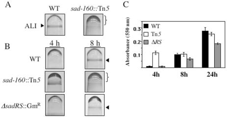
FIG. 1.
Microtiter dish phenotypes of WT strain PA14 and isogenic mutant strains. (A) Biofilm formation phenotypes of the WT and sad-160::Tn5 mutant strains at 8 h in the microtiter dish assay. Cells were grown for 8 h in minimal M63 medium supplemented with MgSO4, glucose, and CAA. The arrowhead indicates crystal violet staining of the biofilm at the ALI. The bracket indicates the increased CV staining below the ALI in the Tn5 mutant. (B) Biofilm formation by the WT, the sad-160::Tn5 transposon mutant, and the ΔsadRS::Gmr mutant strain observed at 4 and 8 h. In panels A and B, the microtiter wells are inverted. (C) Quantification of the biofilm formed by the WT and the sad-160::Tn5 and ΔsadRS::Gmr mutants in microtiter dishes. Biofilms were quantified at 4, 8, and 24 h by solubilization in 30% glacial acetic acid, and the A550 of the resulting solution was measured. See the Materials and Methods for experimental details. Error bars indicate standard deviations.

