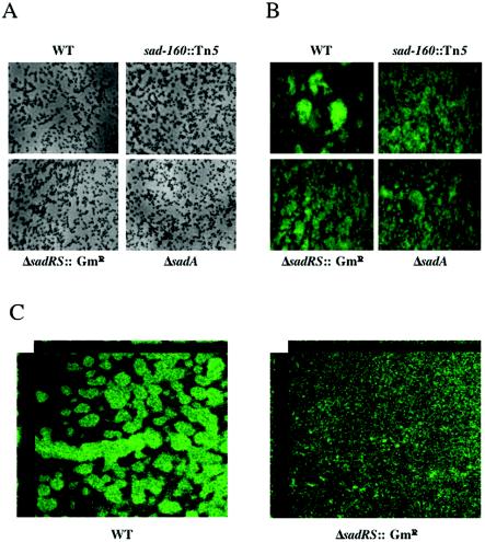
FIG. 3.
Biofilm formation under continuous flow. Biofilm formation at day 2 (A) and day 5 (B) is shown for modified EPRI-grown bacteria in a flow cell. (A) Top-down phase-contrast micrographs at a magnification of ×1,400 are shown for the WT and three representative mutants. (B) Top-down fluorescence images of 5-day-old biofilms at a magnification of ×630. (C) CSLM images of 5-day old biofilms, at a magnification of ×600, of the WT and the ΔsadRS::Gmr mutant, showing the xy and xz planes. Flow cell experiments were performed as described in Materials and Methods.

