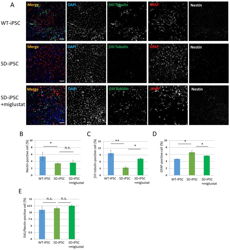
Fig 8. Neural differentiation from NSCs isolated from SDIA-induced colonies.
Primary neurospheres isolated from the SDIA-induced colonies of WT-iPSC and SD-iPSC (with or without miglustat) were dissociated mechanically to single-cell suspensions and replated onto poly-ornithine/fibronectin-coated culture dishes. One hour after plating, the differentiation of NSCs was induced by replacing bFGF-containing medium with fresh N2 medium supplemented with 1% FBS and culturing for 3 days. (A) Immunostaining of differentiated cells for βIII tubulin (green), GFAP (red), and nestin (white) with DAPI nuclear staining (blue). The scale bar indicates 100 μm. (B−D) For each sample, 20 fluorescence images of different fields (1.3 × 1.8 mm) were obtained. The percentages (% of total DAPI count) of NSCs (B), neurons (C), and astrocytes (D) were evaluated. (E) One hour after plating, proliferating NSCs were determined by using the Click-iT EdU Alexa Fluor 488 Imaging kit (green) (S4 Fig). EdU/nestin double-positive cells were counted using the IN Cell Analyzer 2200. Values represent the mean ± S.E. of five independent experiments. n.s.: Not significantly different (P > 0.05), *P<0.05, **P<0.01.

