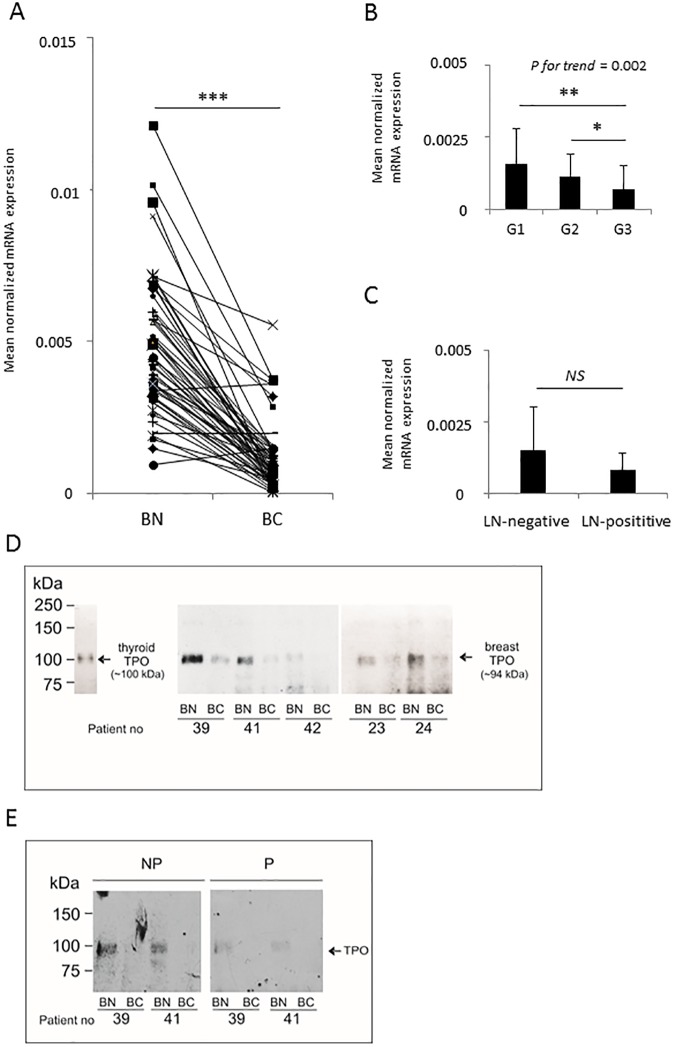
Fig 2. Thyroid peroxidase (TPO) expression in normal and cancerous breast tissue samples in fifty-six patients.
(A) RT-qPCR analysis, (B and C) RT-qPCR analysis of TPO mRNA expression in breast cancer samples according to the cancer grade (B) and the patient nodal status (C). (D-E) TPO protein expression in normal and cancerous breast tissue representative samples by Western blotting. (D) TPO was immunoprecipitated using anti-peptide P14 Ab, and detected with an anti-TPO monoclonal antibody: mAb 47. (E) ab76935 antibody preabsorbed with the excess of highly purified TPO (P) (right panel) or non-preabsorbed ab76935 (NP) (left panel) were used. *: P-value < 0.05; **: P-value < 0.01; ***: P-value < 0.001; NS: not significant; BN: peri-tumoral; BC: breast cancer tissue.

