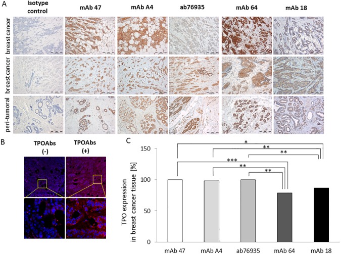
Fig 3. Expression of thyroid peroxidase (TPO) protein in breast tissue samples.
Representative immunostaining results obtained with a panel of monoclonal antibodies (mAbs), (A) and human serum pools (B). Representative images for two breast cancer tissues and one peri-tumoral tissue are shown. Magnification, 100×. (B) Positive staining (red) observed in TPOAb-positive serum pool obtained from patients with autoimmune thyroid disease (AITD; TPOAbs(+)), whereas serum negative for TPOAbs (TPOAbs(-)), used as a negative control, gave weak or no staining. Nuclei (blue) were counterstained with DAPI. Magnification, 200× and 630×. (C) The frequency of TPO positive staining in breast cancer tissue samples (n = 56). In all cases, TPO protein was detected by mAb 47, mAb A4, and ab76935, while mAb 64 and 18 detected TPO in 79% and 87% of all cases, respectively. *: P-value < 0.05; **: P-value < 0.01; ***: P-value < 0.001.

