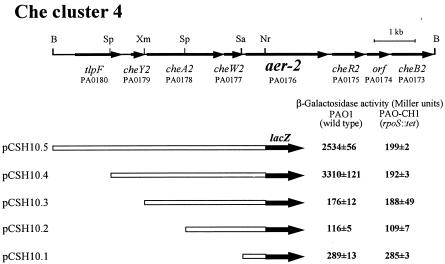
FIG. 2.
Physical map of Che cluster 4 of P. aeruginosa PAO1 (11) and regions cloned in the promoter probe vector pQF50. Specific restriction sites used to isolate each fragment are shown on the map. Restriction sites: B, BamHI; Nr, NruI; Sa, Sau3AI; Sp, SphI; Xm, XmnI. The locations and orientations of tlpF, cheY2, cheA2, cheW2, aer-2, cheR2, PA0174, and cheB2 are indicated by horizontal arrows. Gene identification numbers used in the P. aeruginosa genome sequencing project (http://www.pseudomonas.com/) are indicated below the gene names. Open bars are P. aeruginosa chromosomal DNA fragments subcloned into pQF50. The lacZ gene is shown by the black arrow. β-Galactosidase activities were determined in P. aeruginosa wild-type PAO1 and its rpoS mutant, PAO-CH1, containing the aer-2::lacZ transcriptional fusion plasmids shown. β-Galactosidase activity is shown along with the standard deviation (mean of four independent experiments).

