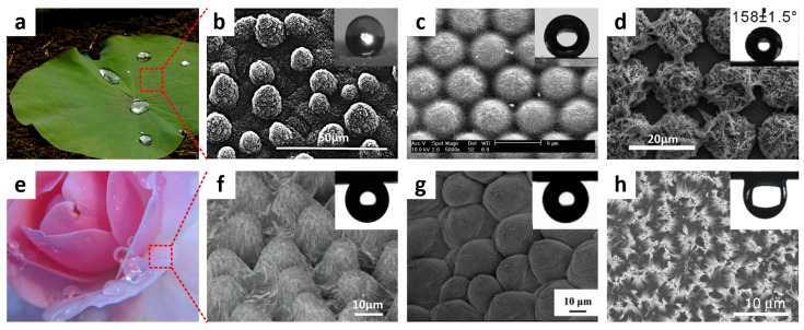
Figure 3.
(a) Image of a lotus leaf (image by GJ Bulte, reproduced under Creative Commons Attribution Share-alike (CC BY-SA) license); (b) the corresponding scanning electron microscopy (SEM) image showing the hierarchical micro/nanostructures comprising papillose cells; (c) SEM image of a microsphere/single-walled carbon nanotube (CNT) composite array; (d) SEM image of a tetragonal array comprising Cu microprotrusions covered by nanostructured Ag dendrites; (e) Photograph of rose petals exhibiting water-adhesive properties; and (f) SEM image of rose petal surface (image by Audrey, reproduced under CC BY license); (g) SEM image of a rose petal-like polystyrene (PS)-film, onto which a water droplet was pinned even when turned upside down; (h) SEM image of Si nanowire arrays; Inset: water droplet deposited on the array after rapid thermal annealing (RTA) at tilt angle of 180°. Reproduced from [32,34,61,62,63,64] with the permission by Springer, Copyright 1997 and by ACS Publications, Copyright 2007, 2008, 2013 and by Elsevier, Copyright 2013.

