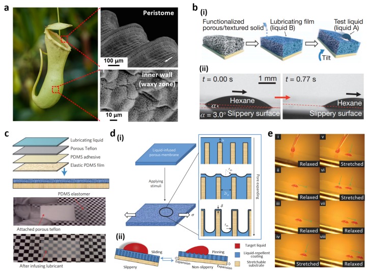
Figure 6.
(a) Image of the Nepenthes pitcher plant (image by William Warby, reproduced under CC BY) and SEM images of the peristome surface and inner wall. The surface presents radial ridges, while the inner wall is covered with waxy crystals; (b) (i) Slippery film fabrication; (ii) Optical micrographs of a sliding hexane drop at a low angle; (c) Porous matrix formation on an elastic PDMS film and photographs of dry and lubricated substrates; (d) (i) Mechanically induced topographical changes in a liquid slippery film upon stretching and (ii) corresponding droplet motions; (e) Sequential photographs of oil droplet movement on the dynamic slippery surface. Reproduced from [13,93,94] with permission by National Academy of Sciences of the USA, Copyright 2004 and by Nature Publishing Group, Copyright 2011, 2013.

