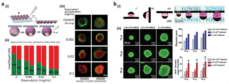
Figure 9.
(a) (i) Hanging drop culture on the indentation-patterned superhydrophobic surface; (ii) Live (green)/dead (red) cell ratios 24 h after the addition of various doxorubicin concentrations for densities of 30,000 (left bar) and 40,000 (right bar) cells/droplet; (iii) Fluorescent images of L929 spheroids in the presence of doxorubicin (ii); (b) (i) Culture medium droplets adhered on the H2-exposed Pd-coated Si NWs for different tile angle (0°, 90°, and 180°) and medium volumes (5, 10, 15 and 20 μL); (ii) Live/dead cell staining, size distribution, and vascular endothelial growth factor (VEGF) protein secretion from spheroids after 4 days of culture at various cell densities and medium volumes. Reproduced from [18,136] with permission by John Wiley and Sons, Copyright 2014.

