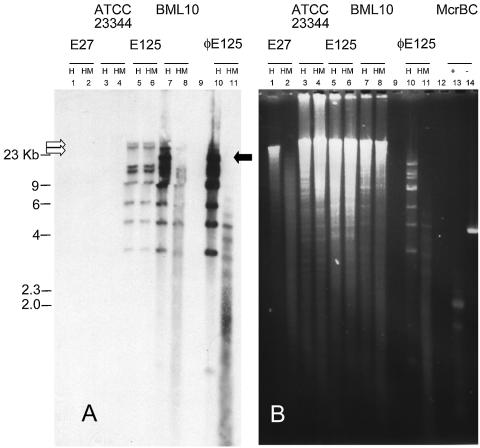
FIG. 1.
Southern blot (A) and electrophoretic (B) analysis of the M.BtaII-induced methylation of ΦE125. (A) Approximately 50 μg of the indicated genomic DNA and ∼10 μg of ΦE125 purified from plate lysates were subjected to digestion by HindIII (H) according to the NEB-specified conditions. (B) Subsequently, half of each reaction mixture was removed and digested with McrBC (HM) under NEB-specified conditions. The DNAs were blotted by using the alkaline transfer protocol of the TurboBlotter (Schleicher & Schuell, Piscataway, N.J.) and photoimmobilized. The ECL probe (ΦE125::HindIII) was synthesized, hybridized, and detected according to the instructions specified by Amersham. The positive controls for McrBC digestion were E27 genomic DNA (lanes 2) and the NEB-supplied positive control (lane 13). The empty arrows indicate the chromosomal junction fragments and the host chromosome, while the filled arrow indicates the fragment that originates from excised viral genomes.

