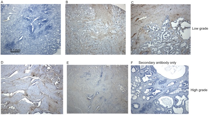
Figure 1.
Fascin-1 staining patterns in human prostate carcinomas. Examples of fascin-1 staining in conventional sections of prostate carcinomas. (A) Uninvolved tissue; (B, C), low/intermediate grade, Gleason score 7 tumours; (D)-(F), high grade, Gleason score 9 tumours. Focal areas of fascin-1–positive tumour cells are indicated by asterisks in (D). All sections were counter-stained with hematoxylin-eosin.

